NCBI Bookshelf. A service of the National Library of Medicine, National Institutes of Health.
Walker HK, Hall WD, Hurst JW, editors. Clinical Methods: The History, Physical, and Laboratory Examinations. 3rd edition. Boston: Butterworths; 1990. Bookshelf ID: NBK366 PMID: 21250207
Definition
Clubbing is a physical sign characterized by bulbous enlargement of the ends of one or more fingers or toes (Figure 44.1). Proliferation and edema of connective tissue result in loss of the normal angle between the skin and nail plate and excessive sponginess of the nail base. Clubbing is usually acquired and is associated with certain cardiopulmonary and gastrointestinal disorders, but may occur in congenital or familial forms. Acropachy is an alternative term for clubbing. Acquired clubbing is often reversible when the associated condition is treated successfully.Hypertrophic osteoarthropathy is an advanced stage of clubbing where there is painful periosteal proliferation of long bones such as the radius, ulna, or tibia. The periostitis is often associated with mild fever and arthralgia or joint effusion. Peripheral neurovascular disease with cyanosis, excessive sweating (hyperhidrosis), paresthesias, and muscular weakness can also occur.
Pachydermoperiostosis is a syndrome that combines hyper-trophic osteoarthropathy and acromegalic features. Patients with this disorder have cylindrical thickening of the limbs due to periostosis and skin changes. The skin changes include marked thickening and oiliness of the skin of the hands, forearms, and legs; accentuation of facial folds; hyperhidrosis of the hands and feet; and reduplication and furrowing of skin over the scalp (called cutis verticis gyrata, or "bulldog scalp"). There may also be macroglossia. The hereditary form of pachydermoperiostosis may be inherited as an autosomal dominant trait with incomplete penetrance and is most severe in males. It usually has its onset in the late teens. The acquired form is not associated with a family history of the disorder, usually occurs in older men, and is associated with coexisting chronic cardiopulmonary or gastrointestinal neoplasm or infection.
Technique
Clubbing
Clubbing is usually painless and bilateral. The patient may be unaware of the gradual enlargement of fingertips or toes until it is brought to his or her attention. Patients with hereditary clubbing may report a family history of clubbing or a long duration of clubbing without any symptoms or signs to suggest an underlying illness. In acute cases, the more rapid onset or mild associated discomfort may draw attention to the deformity. It is important to detect early clubbing because clubbing can be the first clue to a serious underlying disorder that, if treated early, may be curable.Clubbing usually develops first in the thumb and forefinger, and occurs in the other fingers later. It may be difficult to recognize unless the examiner observes the hands (and feet) carefully. Two signs are characteristic of early clubbing: the "floating nail" sign and the "profile" sign.
The "floating nail" sign is easily demonstrated (Figure 44.2). Normally, the root of the nail plate lies snugly against the bone of the distal phalanx; pressure on the root produces no movement. With clubbing, the root is separated from bone by connective tissue and edema; pressure upon the nail plate moves it toward the bone. The base of the nail becomes resilient and springy, and the nail feels as if it is floating on a cushion. As clubbing progresses, the nail becomes loosely attached, and the free edge of the nail plate may become visible or palpable as a horizontal ridge over the dorsal aspect of the finger.
The "profile" sign is illustrated in Figures 44.2 and 44.3. Normally, the angle between the nail plate and the skin overlying the proximal part of the distal phalanx is about 160 degrees or less. With clubbing, proliferation of tissue under the nail plate causes this angle to increase to more than 160 degrees. In fact, the angle may be entirely lost and the nail plate and skin lie in a straight line (180-degree angle). As clubbing progresses, the angle exceeds 180 degrees. Eventually, the profile of the fingertip becomes bulbous.
Obliteration of the angle between the nail and the nailbed is the first sign of clubbing and its most constant feature; indeed, many require it for the diagnosis. Because it can be easily detected and precisely defined, the examiner should rely on this sign when uncertain if a patient's fingers are truly clubbed.
Early clubbing must be distinguished from several other abnormalities of the fingertip. These include: (1) increased nail curvature, a normal finding in many adults (especially blacks); (2) chronic paronychiae, in which the soft tissue at the base of the nail is inflammed and swollen; and (3) felons or abscesses of the terminal pulp space, in which there is swelling over the palmar surface of the fingertip. In none of these conditions, however, is the fundamental angle between nail and terminal phalanx altered. A simple maneuver (described by Schamroth) readily differentiates true clubbing from these pseudo-clubbing conditions. The examiner asks the patient to hold the two thumbs back to back with the two fingertips and interphalangeal joints aligned. Because the base angle is unaltered in these conditions, an elongated, diamond-shaped area of empty space is apparent. In true clubbing, this empty space is lost. (Schamroth, while himself ill with subacute bacterial endocarditis, noted this finding as one of the earliest signs of clubbing; he also reported that the window of empty space reappeared 2 months after his endocarditis had been treated successfully.)
Increased nail curvature, when it occurs in clubbing, appears later than either proliferation of tissue at the nail base or loss of the normal angle between distal phalanx and nail. Though increased nail curvature is not specific to clubbing, when it occurs, the entire nail assumes an abnormally convex appearance (often described as the "watch-crystal" nail). Later still, when the entire distal phalanx is enlarged and bulbous, it may resemble a drumstick.
Clubbing of the toes is more difficult to recognize because the tips of the toes may normally appear somewhat bulbous. True clubbing is usually easiest to recognize in the great toe. In addition, the examiner may learn to recognize it because clubbing of the toes is frequently found in patients with clubbing of the fingers. Finally, in acute cases, the patient may have noted either mild discomfort or changed appearance of the digits.
Hypertrophic Osteoarthropathy
Hypertrophic osteoarthropathy should always be sought when clubbing is found. The symptomatic patient with hypertrophic osteoarthropathy will complain of severe, burning, deep pain over the distal extremities, especially of the legs, that is worsened at night and on dependency. In early cases, the examiner may note warmth, reddening, or brawny edema of the skin over the distal long bones (particularly over the shins). In acute or severe cases, there may be exquisite tenderness of the feet and legs and the hands and forearms, and tenderness and effusions of the adjacent joints (the ankles, knees, wrists, and fingers). The examiner may elicit tenderness over bony prominences by simply pressing or by encircling the limb (e.g., the wrist) with his own fingers like a bracelet and gently squeezing it. Later, autonomic changes such as flushing, sweating, or blanching (Raynaud's phenomenon) may be noted in the affected hands and feet.Pachydermoperiostosis
Patients with pachydermoperiostosis most often complain about their changing appearance or the excessive perspiration, especially of the hands and feet. During the active phase of their disorder, they may have mild bone pain or arthralgia. Later, they may complain of clumsiness of their fingers, ptosis from thickening of the skin over their eyelids, or facial palsy from thickening of the skull. On examination of the skeleton, there is clubbing of the fingers and toes, accompanied by "spadelike" or "pawlike" enlargement of the hands and feet; joint effusions may be present. The skin changes include excessive sweating, generalized thickening (called pachyderma) and redundancy, especially over the forehead and scalp, leading to characteristic "bulldog" furrowing (cutis verticis gyrata) and leonine facies, sometimes mistaken for leprosy. The cylindrical thickening of the extremities resulting from a combination of bony enlargement and thickening of the skin is often the most impressive physical finding and may suggest the diagnosis of acromegaly.Further Evaluation
In all three conditions (i.e., clubbing, hypertrophic osteoarthropathy, and pachydermoperiostosis), the physician should specifically examine for findings of associated illness, including wheezes, rales, pleural effusion, supraclavicular adenopathy, cardiac murmurs, Roth spots, peripheral stigmata of endocarditis, splenomegaly, jaundice, vascular angiomata, palmar erythema, hepatomegaly, abdominal mass (suggesting regional enteritis), thyromegaly, and ophthalmopathy.In addition, because clubbing may be a manifestation of either hypertrophic osteoarthropathy or pachydermoperiostosis, the clinician may want to obtain x-rays of the distal extremities. In early clubbing, x-rays will be normal except for increased soft tissue. In the later stages, there may be fraying of the edges of the distal phalanx, followed by gradual resorption of the bone (acro-osteolysis). X-rays of patients with hypertrophic osteoarthropathy may show symmetrical, irregular periosteal proliferation, calcification and new bone formation (periostitis) over the distal arms and legs, or acro-osteolysis of the distal phalanges of fingers and toes. The finding that 99m-technetium pyrophosphate bone scans are more sensitive than plain x-rays in detecting early periostitis can be helpful in difficult cases. In advanced cases of osteoarthropathy, and in pachydermoperiostosis, x-rays may show extreme degrees of periostitis in almost any bone, although the skull, vertebrae, and articular surfaces are usually spared.
Synovial fluid obtained by arthrocentesis in patients with painful joint effusions in osteoarthropathy are usually "non-inflammatory," with a leukocyte count less than 2000/mm3 and with 0 to 50% neutrophils. Interestingly, this fluid tends to clot spontaneously after aspiration, a finding usually reserved for inflammatory effusions.
Further radiologic or laboratory studies, such as a chest x-ray, small bowel series, thyroid function panel, or blood cultures, may be performed to examine for underlying illness. The patient's symptoms and signs will indicate the appropriate procedures.
Basic Science
Although many explanatory theories have been put forward, the etiology of clubbing is unknown. Flavell (1956) quotes West, who said in 1907 that "clubbing is one of those phenomena with which we are all so familiar that we appear to know more about it than we really do."Shneerson (1981) reviews the four most likely mechanisms underlying clubbing. They include a circulating vasodilator, tissue hypoxia, a neurocirculatory reflex, and genetic factors. For acquired clubbing, the most likely mechanism postulates a neurocirculatory reflex, leading to increased blood flow through multiple arteriovenous shunts in the distal phalanges. Increased blood flow then leads to tissue hypertrophy and hyperplasia on a nutritional basis. The evidence for this mechanism is threefold: (1) clubbing may resolve after vagotomy; (2) disturbances of the vasculature such as arteriovenous fistulae and aneurysms are associated with its development; and (3) Racoceanu (1971) and others have demonstrated increased blood flow to digitial capillaries in acquired clubbing.
Flavell (1956) first proposed the neurocirculatory reflex theory to explain hypertrophic osteoarthropathy, after he observed that severing the vagus nerve could reverse it even when the underlying lung cancer was unresectable. According to this theory, there is a reflex in which afferent impulses travel by the vagus nerve from the inciting focus (such as a lung tumor) to the central nervous system. The efferent limb of the proposed reflex is unknown, but presumably some humoral substance or neural impulse mediates the vascular changes, leading to hypertrophy of the fibroconnective tissues. Others have supported such a mechanism by observations that the visceral organs in which associated diseases occur are all innervated by the glossopharyngeal or vagus nerves.
Strong support for this theory has also come from a study by Gold and colleagues (1979). They studied a unique case of unilateral clubbing secondary to a posttraumatic aneurysm of the ulnar artery, which resolved after resection of the aneurysm. Using the contralateral hand of the otherwise healthy patient, they conducted detailed studies of the microcirculation by Doppler flow recordings, differential pulse pressure measurements, angiography, measurements of reactive hyperemia, and differential capillary blood gas determinations. They interpreted their findings of increased blood flow, decreased peripheral resistance, and increased oxygenation to support the neurocirculatory reflex mechanism. They postulated that the efferent limb of the reflex was cholinergic fibers of the autonomic sympathetic innervation of digital arteriovenous shunts.
However, different mechanisms may cause acquired and hereditary clubbing. Some workers have found that digital capillary blood flow, measured by washout of krypton-85 solution, is increased in acquired clubbing but normal in hereditary clubbing. An alternative explanation for this finding may be that in hereditary clubbing the condition has been studied only after the condition has stopped progressing; the physiologic findings might be similar in the active phase.
There may be different pathogeneses of acquired clubbing and osteoarthropathy. Each may occur without the other. Some conditions frequently associated with clubbing are seldom seen with osteoarthropathy, and vice versa. Left-to-right pulmonary arteriovenous shunting may be important in the pathogenesis of osteoarthropathy in some patients, such as those with chronic liver disease, but cannot explain others, such as those with marked right-to-left shunts. Overall, however, both the pathology and physiology in limbs affected by osteoarthropathy resemble that of the digits affected by clubbing.
Study of synovial tissue from patients with hypertrophic pulmonary osteoarthropathy has shown evidence of microvascular injury, perhaps related to electron-dense deposits found in the synovial vessel walls. These dense deposits do not stain for immunoglobulins or complement, however, and may simply represent proteins such as fibrinogen that have leaked through the damaged blood vessel walls.
The pathogenesis of pachydermoperiostosis seems quite different. Blood flow in the limbs as measured by plethysmography and arteriovenous oxygen differences is normal, capillaroscopy shows few vessels, and angiograms show slowed flow with segmental arterial narrowing. Various humoral agents such as hormones have been proposed to explain its pathogenesis, but there is little evidence to support such a mechanism. For example, despite the acromegaloid features of affected patients, there has been no evidence of ectopic secretion of excessive amounts of growth hormone.
Clinical Significance
The earliest known description of clubbing is that of Hippocrates, who noted in the fifth century b.c. its association with chronic empyema. He wrote: "The nails of the hand are bent; the fingers are hot especially in their extremities."Clubbing and hypertrophic osteoarthropathy are important to recognize because they are usually associated with serious underlying disease. In addition, because clubbing may precede any other evidence of the underlying disorder, it may serve as the only clue to an otherwise silent lesion. It may, for example, allow early recognition and surgical cure of an otherwise asymptomatic lung tumor.
Review of the literature indicates that between 75 and 80% of cases are associated with chronic pulmonary diseases; 10 to 15% are associated with cardiovascular diseases; 5 to 10% with chronic hepatic and gastrointestinal disorders; and 5 to 10% with miscellaneous disorders.
Pulmonary disease is the most common cause (Table 44.1). Primary or secondary malignancy of the bronchus or lung, as well as tumors of the pleura, mediastinum, thymus, and chest wall, are perhaps the most worrisome associations. But a variety of chronic pulmonary infections, including lung abscess, chronic empyema, pneumoconiosis, chronic pneumonitis, cystic fibrosis, emphysema with chronic suppuration, actinomycosis, and bronchiectasis, can also produce the characteristic picture. Clubbing is said to be uncommon in uncomplicated pulmonary tuberculosis, but may occur in up to 25% of cases with supervening bronchiectasis or chronic cavitation. More rarely, it has been described in patients with chest deformities, aortic aneurysm with compression of lung, and neurogenic tumors of the diaphragm.
Cardiovascular diseases rank second among the causes of clubbing and hypertrophic osteoarthropathy (Table 44.1). Associated diseases include cyanotic congenital heart diseases (diseases with right-to-left shunts), subacute bacterial endocarditis, chronic congestive heart failure, cor pulmonale, cardiac tumors, pulmonary arteriovenous fistulas, and secondary polycythemia.
Chronic hepatic and gastrointestinal disorders associated with clubbing include primary and secondary biliary cirrhosis, amebic liver abscess, hepatic amyloidosis, ascariasis, malaria, intestinal tuberculosis, chronic amebic dysentery, ulcerative colitis, regional enteritis, sprue, intestinal polyposis, abdominal lymphoma, and carcinomas of the nasopharynx, esophagus, stomach, and colon (Table 44.1).
Miscellaneous associations include thyrotoxicosis (thyroid acropachy), hyperparathyroidism, pregnancy, chronic osteomyelitis with amyloidosis, chronic pyelonephritis, pseudohypertrophic muscular dystrophy, hemoglobinopathies, chronic mountain sickness (Monge's disease), syringomyelia, renal carcinoma, chronic myelocytic leukemia, and exposure to toxins such as arsenic, mercury, silica, beryllium, phosphorus, and alcohol (Fable 44.1).
Asymmetrical clubbing usually indicates impaired regional blood flow caused by localized vascular disease. Unilateral clubbing in the upper extremity may be due to anomalies of the aortic arch, aortic or subclavian artery aneurysm, pulmonary hypertension with patent ductus arteriosus, and brachial arteriovenous aneurysm or fistula. Rarely, it has been described with recurrent dislocation of the shoulder and superior sulcus (Pancoast) tumor. Clubbing of toes but not fingers suggests coarctation of the aorta. Unidigital clubbing is seen following median nerve injury and, rarely, with sarcoidosis. Recurrent clubbing may occur during pregnancy in otherwise healthy women. Familial clubbing is of no clinical import (unless associated with pachydermoperiostosis).
References
- Booth BW, Van Nostrand D, Graeber GM. Hypertrophic pulmonary osteoarthropathy and breast cancer. So Med J. 1987;80:383–86. [PubMed: 3824029]
- Epstein O, Ajdukiewicz AB, Dick R. et al. Hypertrophic hepatic osteoarthropathy: clinical, roentgenologic, biochemical, hormonal and cardiorespiratory studies, and review of the literature. Am J Med. 1979;67:88–97. [PubMed: 463921]
- Fischer DS, Singer DH, Feldman SM. Clubbing, a review, with emphasis on hereditary acropachy. Medicine. 1964;43:459–77. [PubMed: 14183519]
- Flavell G. Reversal of pulmonary hypertrophic osteoarthropathy by vagotomy. Lancet. 1956;1:260–62. [PubMed: 13287123]
- Gold AH, Bromberg BE, Herbstritt JG. et al. Digital clubbing: a unique case and a new hypothesis. J Hand Surg. 1979;4:60–65. [PubMed: 759505]
- Herbert DA, Fessell WJ. Idiopathic hypertrophic osteoarthropathy (pachydermoperiostosis). West J Med. 1981;134:354–57. [PubMed: 7245739]
- Herman MA, Massaro D, Katz S. et al. Pachydermoperiostosis—clinical spectrum. Arch Intern Med. 1965;116:918–23. [PubMed: 5848228]
- Holling HE, Brodey RS, Boland HC. Pulmonary hypertrophic osteoarthropathy. Lancet. 1961;2:1269–74. [PubMed: 13908437]
- *Lipman BS, Massie E. Clubbed fingers and hypertrophic osteoarthropathy. In: MacBryde CM, Blacklow RS, eds. Signs and symptoms: applied pathologic physiology and clinical interpretation. Philadelphia: J. B. Lippincott, 1970;256–71.
- Pyke DA. Finger clubbing: validity as a physical sign. Lancet. 1954;2:352–54. [PubMed: 13184677]
- Racoceanu SN, Mendlowitz M, Suck AF. et al. Digital capillary blood flow in clubbing85Kr studies in hereditary and acquired cases. Ann Intern Med. 1971;75:933–35. [PubMed: 4944158]
- Rosenthall L, Kirsch J. Observations on radionuclide imaging in hypertrophic pulmonary osteoarthropathy. Radiol. 1976;120:359–62. [PubMed: 935484]
- Schamroth L. Personal experience. S Afr Med J. 1976;50:297–300. [PubMed: 1265563]
- Schumacher HR. Articular manifestations of hypertrophic pulmonary osteoarthropathy in bronchogenic carcinoma. Arthr Rheum. 1976;19:629–36. [PubMed: 938592]
- Segal AM, Mackenzie AH. Hypertrophic osteoarthropathy: a 10-year retrospective analysis. Semin Arthr Rheum. 1982;12:220–32. [PubMed: 6101214]
- Shneerson JM. Digital clubbing and hypertrophic osteoarthropathy: the underlying mechanisms. Br J Dis Chest. 1981;75:113–31. [PubMed: 7023525]
- Vogl A, Goldfischer S. Pachydermoperiostosis: primary idiopathic hypertrophic osteoarthropathy. Am J Med. 1962;33:166–87. [PubMed: 13926461]
Figures
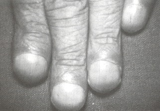
Figure 44.1
Clubbing of the fingers of one hand.
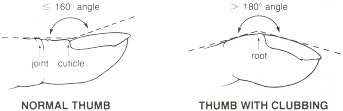
Figure 44.2
The normal and clubbed fingertip. The "floating nail" sign is demonstrated by applying pressure at the point indicated as the root of the nail plate. Normally, pressure there produces no movement. With clubbing, there is movement toward the bone. The "profile" sign is produced by an increased angle between the nail plate and the skin overlying the proximal part of the distal phalanx. Normally, the angle is about 160 degrees or less. As clubbing progresses, the angle exceeds 180 degrees.
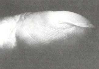
Figure 44.3
The clubbed fingertip in profile.
Tables
Table 44.1Conditions Associated with Clubbing
| I. Symmetrical Clubbing |
 Pulmonary disease Pulmonary disease |
| Neoplasm |
| Bronchogenic carcinoma |
| Pleural mesothelioma |
| Thymoma |
| Metastatic carcinoma |
| Chronic infection |
| Lung abscess |
| Chronic empyema |
| Pneumoconiosis |
| Chronic pneumonitis |
| Cystic fibrosis |
| Emphysema with chronic suppuration |
| Actinomycosis |
| Bronchiectasis |
| Cardiovascular disease |
| Cyanotic congenital heart disease |
| Subacute bacterial endocarditis |
| Cor pulmonale |
| Secondary polycythemia |
| Chronic congestive heart failure |
| Cardiac tumors |
| Pulmonary arteriovenous fistula |
| Arterial graft infection |
| Hepatic and gastrointestinal disease |
| Hepatic |
| Cirrhosis (all types) |
| Amebic abscess |
| Amyloidosis |
| Gastrointestinal |
| Chronic infection |
| Ascariasis |
| Malaria |
| Intestinal tuberculosis |
| Chronic amebic dysentery |
| Chronic inflammation |
| Ulcerative colitis |
| Regional enteritis |
| Sprue |
| Neoplasm |
| Intestinal polyposis |
| Abdominal lymphoma |
| Nasopharyngeal carcinoma |
| Esophageal carcinoma |
| Gastric carcinoma |
| Colonic carcinoma |
| Miscellaneous |
| Endocrine conditions |
| Hyperthyroidism |
| Hyperparathyroidism |
| Pregnancy |
| Chronic infections |
| Chronic osteomyelitis with amyloidosis |
| Chronic pyelonephritis |
| Congenital disorders |
| Muscular dystrophy |
| Hemoglobinopathies |
| Idiopathic disorders |
| Chronic mountain sickness |
| Syringomyelia |
| Neoplasms |
| Renal carcinoma |
| Chronic myelocytic leukemia |
| Toxin exposure |
| Arsenic |
| Mercury |
| Silica |
| Beryllium |
| Phosphorus |
| Alcohol |
| Chronic laxative abuse |
| Familial |
| II. Asymmetrical Clubbing |
| Unilateral |
| Anomalous aortic arch |
| Aortic or subclavian artery aneurysm |
| Pulmonary hypertension with patent ductus arteriosus |
| Brachial arteriovenous aneurysm or fistula |
| Recurrent shoulder dislocation |
| Superior sulcus (Pancoast) tumor |
| Unidigital |
| Median nerve injury |
| Sarcoidosis |
| Clubbing of toes without fingers |
| Coarctation of aorta |
Copyright © 1990, Butterworth Publishers, a division of Reed Publishing.
The function of thyroid gland is to produce thyroid hormone in presence of iodine. The production rate of thyroid hormone is very delicate, as under production (hypothyroidism) and even over production (hyperthyroidism) causes thyromegaly. Read More At http://healthsaline.com/thyromegaly.html
Trả lờiXóa