We were never tired of visiting and examining and ausculting, and of examining and ausculting again and again.
—Peter Mere Latham (1789–1875)
A bruit is an audible vascular sound associated with turbulent blood flow. Although usually heard with the stethoscope, such sounds may occasionally also be palpated as a thrill. In the head and neck, these auscultatory sounds may originate in the heart (cardiac valvular murmurs radiating to the neck), the cervical arteries (carotid artery bruits), the cervical veins (cervical venous hum), or arteriovenous (AV) connections (intracranial AV malformations). These sounds may be normal, innocent findings (i.e., a venous hum in a child) or may point to underlying pathology (i.e., a carotid artery bruit caused by atherosclerotic stenosis in an adult). Head and neck bruits loom especially important today because physicians encounter arterial occlusive disease more frequently as a greater proportion of our population lives longer.
What are the clinical circumstances where head and neck auscultation is indicated? Evaluation often begins with patient symptoms related to cardiovascular or neurologic disease. This involves a through, directed physical examination following pertinent historical information. Second, the necessity for head and neck auscultation may be part of repeated or sequential physical examinations directed by new data from subsequent history, physical examination, or laboratory information obtained later in the hospital course. Finally, auscultation of head and neck vascular sounds may be part of a routine, complete (comprehensive) physical examination, especially in subsets of patients at risk for neurologic or cardiovascular disease.
Cranial and orbital bruits are vibrations resulting from turbulence in intracranial or extracranial vessels. Although usually systolic in timing, these bruits may extend into diastole or even be continuous. These sounds may originate within the cranium or be transmitted from arteries in the neck or, occasionally, from cardiac valvular lesions. The orbits provide relative "windows" for transmission of intracranial sounds, with minimal bony dissipation. Indication for cranial and orbital auscultation usually follows from historical physical examination or laboratory evidence of cranial—cervical disorders such as seizures, headaches, stroke syndromes, intracranial mass lesions, or carotid bruits.
Neck auscultation is commonly indicated for initial evaluation of stenotic or embolic cerebrovascular symptoms, or as part of a comprehensive physical examination in asymptomatic patients at risk for atherosclerosis. Cervical bruits and hums may arise from neck arteries or veins, and may be innocuous findings or indicate underlying pathology. Bruits arising in the carotid arteries are produced by intrinsic stenosis or, occasionally, with vascular occlusion from extrinsic compression. Depending on a variety of factors, these bruits may be systolic, primarily systolic with extension into diastole, or continuous. The cervical venous hum is auscultated over the internal jugular veins in many normal children. Commonly a continuous high-pitched sound, it is occasionally more prominent in diastole. It occurs more frequently on the right than on the left, and may be present bilaterally.
Supraclavicular bruits during systole are a frequent finding in normal children and in adults with subclavian or vertebral artery stenosis. Supraclavicular auscultation is usually initiated to evaluate vertebral artery occlusive symptoms, arm claudication, or "subclavian steal" in the adult with atherosclerosis.
By the management of pressure with a stethoscope over or near large veins, the venous murmur may often be raised, by a gradual swelling, into a more or less musical hum . . . I propose to denominate this "venous hum."In physical evaluation of these sounds, an important aspect is their relationship to the cardiac cycle. Equally important and useful is an appreciation of their dynamic quality—that is, findings may change from time to time in the same patient depending on a variety of factors (e.g., activity, stress, fever). From a physical diagnosis standpoint, this dynamic characteristic may be exploited by use of maneuvers designed to alter these vascular sounds in predictable ways.
—James Hope (1801–1841)
Auscultation should take place in a quiet room with both patient and examiner relaxed and comfortable. Although it is often stated that what one puts in the ears is of less importance than what lies between them, an efficient, well-designed stethoscope is indispensable!
Cranial bruits should be listened for over the skull and eyeballs, the latter location being the most favorable for fainter sounds. The sitting patient is asked to close both eyes gently, and the examiner, facing the patient, places the stethoscope bell firmly over one closed eye. The examiner's other hand (thumb) rests lightly on the contralateral carotid artery for timing purposes and for steadying the head and neck (Figure 18.1). The patient is then asked to open the other eye and look at a fixed object, as this helps eliminate eyelid tremor. Finally, the patient is asked to hold his breath. Orbital bruits may be faint and high-pitched, and the examiner should concentrate especially on the systolic phase of the cardiovascular cycle. After orbital examination, the patient's skull is auscultated with the bell over the mastoid, frontal, and parietal areas. Auscultation is then carried out over the other eye and hemicranium in a similar manner, comparing symmetrical regions if indicated. The examiner should allow the patient to resume respirations after each auscultation.

Figure 18.1
Technique for orbital auscultation.

Figure 18.2
Arrows indicate points of manual compression of the right superficial temporal and facial arteries.
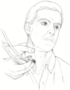
Figure 18.3
Technique for ausculting and abolishing venous hum.
Arterial tortuosity and kinking, which may be palpated in the cervical region, can be a source of turbulence and bruits. Also, compression syndromes, such as the cervical rib or scalenus anticus syndrome, may also be manifest by bruits in the neck. The compression and resulting bruit may be increased by having the patient brace the shoulders back and inspire deeply while the examiner exerts mild downward traction on the homolateral arm (Adson's maneuver). The examiner may simultaneously palpate the radial pulse, which should diminish during this maneuver.
Supradavicular auscultation is performed while the patient sits looking forward, with the shoulders relaxed and hands resting on the lap. The stethoscope bell is lightly applied in each supraclavicular fossa over the subclavian artery. As usual, the examiner's free hand palpates the contralateral carotid pulse for timing purposes. If a bruit is appreciated, firmly compress the patient's ipsilateral radial artery, noting the effect on the murmur. Finally, with the patient supine, continue auscultation after he or she repeatedly squeezes a rubber ball or other pliable object for 30 to 60 seconds. The patient is supine because such exercise may provoke syncope or arm pain while altering the observed bruit.
Whatever constricts an orifice, whatever dilates a cavity, whatever establishes an orifice or cavity where none shall be, will disturb the even flow of blood and produce vibrations and a murmur.Interpretation of vascular sounds in any anatomic location requires a basic understanding of vascular hemodynamics. An arterial bruit usually implies stenosis at or proximal to the site of auscultation. Very severe obstruction, however, may not manifest a bruit; conversely, bruits may be heard over unoccluded normal arteries in certain high-flow circumstances. Because a bruit may be auscultated directly over a stenosis, or distally in the direction of the blood jet producing the vibrations, it is important to trace proximally the audibility of an arterial sound to determine the exact anatomic site of the flow disturbance.
—Samuel Jones Gee (1839–1911)
Intraarterial pressure/flow relationships are approximated as follows:

For any instantaneous stenotic flow:

Additional extrastenotic factors operating in most regional vascular beds alter flow and pressure and bruit characteristics for any fixed stenotic lesion. These factors may be divided into four categories: inflow factors, side branch factors, collateral vessel development, and outflow factors (Figure 18.4). The presence or absence of these variables can affect bruit features and often can be altered for diagnostic purposes. From a diagnostic viewpoint, maneuvers accentuating or localizing bruits are helpful during physical examination. Awareness of all factors influencing pressure and flow across a stenosis thus allows estimation of degree and/or location of that stenosis.
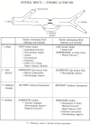
Figure 18.4
Summary of factors altering intensity and duration of arterial bruits. See text for details. A-V = arteriovenous; CHF = congestive heart failure. From Kurtz KJ. Dynamic vascular auscultation. Am J Med 1984;76:1066–74. Reproduced with permission. (more...)
In the second category (side branch factors), compression of arterial side branches should augment a bruit in the main artery and diminish a side branch bruit, whereas augmentation of flow through a side branch would have the opposite effect. This concept is especially applicable for evaluation of carotid artery bruits.
The extent of collateral circulation around a critical stenosis influences the characteristics of a bruit. With internal carotid artery stenosis, for example, collateral formation is usually limited, and the character and timing of a bruit correlate rather closely with the degree of stenosis. In contrast, in the lower extremity, extensive collateral circulation may develop around a stenosis, thus altering the gradient across that stenosis and obscuring the degree of narrowing.
Outflow factors are often important in vascular auscultation. Arteriolar vasodilatation caused by regional exercise is valuable for accentuating bruits in the extremities.
These principles apply directly to auscultation of cranial, cervical, and supraclavicular vessels. The aortic arch arteries are a common site of atherosclerotic lesions, 80% of which arise at the bifurcation of the common carotid arteries. Detection of internal carotid lesions is obviously important, but 10% of carotid bruits originate in the external carotid arteries, representing little risk of subsequent stroke.
Side branch factors can be dynamically altered during cervical auscultation to accentuate and/or localize a carotid bruit. After carotid auscultation at rest, the examiner compresses the superficial temporal and facial arteries arising from the ipsilateral external carotid artery (Figure 18.2). An internal carotid bruit may become louder and longer, whereas an external carotid bruit generally becomes noticeably shorter and softer with compression.
Internal carotid outflow factors may be altered during cervical auscultation. Carbon dioxide is a powerful vasodilator of normal cerebral vessels. Holding the breath raises arterial carbon dioxide tension, causing active dilatation of cerebral arterioles and augmenting blood flow through intracerebral arterioles. Breath holding ordinarily causes a 30% increase in internal carotid bruit intensity, whereas a decrease occurs in external carotid murmurs. Internal carotid outflow also increases with careful compression of the contralateral common carotid artery during auscultation. This compression decreases flow through the circle of Willis, with intensification and prolongation of an internal carotid bruit, and diminution of an external carotid murmur (see Table 18.1 for summary).
Artenovenous fistulas and malformations shunt large volumes of blood at rapid flow from high-pressure arteries into low-pressure veins, producing a continuous murmur with systolic accentuation.
The turbulence of the venous hum, heard over the internal jugular veins, is caused by the combined effects of gravity on blood flow and partial compression of the internal jugular vein by the transverse process of the atlas. This hum occurs more frequently on the right side, partly because the right internal jugular and innominate veins provide short, straight access to the superior vena cava, thus increasing flow velocity in the right jugular venous return. A Valsalva maneuver, manual compression of the internal jugular vein, and the recumbent position all decrease venous flow and obliterate this sound. In contrast, conditions such as upright posture, rapid atrial filling (diastole), and inspiration all increase jugular venous flow and accentuate a venous hum.
In supraclavicular auscultation, arm exercise has been utilized to increase bruits caused by stenosis in a subclavian artery. Local exercise of an extremity supplied by a stenotic artery should increase peripheral outflow, producing greater stenotic turbulence and augmenting a bruit. In contrast, ipsilateral radial artery compression, which decreases subclavian outflow, should shorten or obliterate a subclavian murmur.
Table 18.1Dynamic Maneuvers Useful in Auscultation of Carotid Artery Bruits
- Note: ↑ ↓ indicates change in pitch, intensity, length of bruit.
The trained ear is probably still the best instrument for recognition of most murmurs.After a vascular sound is auscultated in the head, neck, or supraclavicular fossa, the examiner must determine the significance of the sound. Is it nonorganic (innocent), pointing to no regional anatomic disease? Or is it organic (pathologic), indicating localized vascular obstruction and need for further evaluation?
—Abe Ravin (1909–1978)
Table 18.2Conditions Frequently Associated with Head Bruits
Cranial and Orbital Bruits
Cranial and orbital bruits (Table 18.2) are usually normal, innocent findings in younger persons, occurring in 30 to 60% of normal infants and children under 6 years of age. In adults, nonorganic orbital bruits may be noted in patients with high cardiac output states, or those with decreased blood viscosity, as with severe anemia. Often systolic in timing and auscultated over both orbits, these bruits are especially frequent in patients with several simultaneous reasons for increased vascular flow, such as anemia and high fever.If the above conditions are excluded, cranial-orbital bruits in the adult are usually organic (pathologic) and indicate an underlying abnormality. Arteriovenous fistulas, arteriovenous malformations, and intracranial hemangiomas are associated with a prominent bruit in 30 to 50% of cases (Table 18.2). Often these are continuous bruits and are appreciated best over the orbits, where osseous dissipation of vibrations is minimized. Atherosclerosis of the arteries in the neck may rarely produce an orbital bruit. With tight internal carotid stenosis, either extracranially or intracranially at the carotid siphon, a bruit may radiate cephalad and be auscultated over the ipsilateral orbit. With total unilateral carotid occlusion, there may be no bruit over the affected carotid and increased flow through the unoccluded carotid, which can cause a soft bruit over the contralateral eyeball (Table 18.3).
Other intracranial pathology affecting vascular structures may produce soft systolic orbital bruits (Tables 18.2 and 18.3). Positive ausculatory findings should suggest these diagnoses only if the entire clinical picture is supportive.
Cervical Bruits
As suggested previously, nonorganic (innocent) vascular sounds are common in the cervical region and obviously must be differentiated from organic (pathologic) bruits. Children and adults with high cardiac output often have innocent cervical bruits related to high carotid flow. The venous hum, common in normal children, is less frequent in adults except in high cardiac output states. If especially loud in young persons, the hum may radiate to the first and second intercostal spaces and be misdiagnosed as a patent ductus arteriosus if heard on the left.Table 18.3Conditions Occasionally Associated with Head Bruits
All the above ausculatory entities can be differentiated from carotid bruits by clinical techniques discussed previously. Another cause of neck bruits unrelated to carotid pathology is the cervical bruit of thyrotoxicosis (see Chapter 135). In this condition, blood flow through the enlarged thryoid gland increases five- to tenfold, producing a systolic bruit directly over the thyrotoxic goiter. One must distinguish this bruit from a venous hum or an innocent high-flow carotid bruit, findings also present in severe thyrotoxicosis.
If the above nonorganic conditions are eliminated, a cervical bruit should raise the question of carotid artery stenosis with its associated risk of ischemic stroke. Eight million adults in this country have carotid bruits, one-third of whom have experienced focal ischemic symptoms. The Joint Study of Extracranial Arterial Occlusion estimates that 75%) of patients with ischemic stroke syndromes have at least one obstructive lesion at a surgically accessible site. The usual cause of cervical artery obstruction is atherosclerosis.
The exact role of vascular surgery in patients with truly asymptomatic bruits, and in those who have bruits with transient ischemic symptoms or bruits with previous cerebral infarction, continues to be debated vigorously. Nevertheless, diagnosis begins in the office or at the bedside. Even a "silent" neck may harbor advanced atherosclerotic disease because bruits are not present over total carotid occlusion, or when residual lumen diameter is less than 1 mm. But otherwise, cervical auscultation for carotid-associated symptoms of central nervous system disease can localize and grade a stenotic lesion producing transient ischemic attacks (TIAs). With increasing occlusion, an internal carotid bruit becomes higher pitched, more intense, and longer in duration, even extending into diastole with very tight stenosis. Moreover, dynamic maneuvers may accentuate a bruit caused by a plaque narrowing the artery only minimally, but still producing TIAs by microembolization. Or, if a carotid bruit can be localized to the external carotid artery (10%), no further evaluation is usually required; however, an asymptomatic internal carotid bruit should at least be followed closely. Even a truly asymptomatic carotid bruit may warn of future morbidity from atherosclerotic disease in other parts of the body because evidence of atherosclerotic disease in one region often indicates significant lesions in other vascular areas.
A vascular history suggesting stenotic or embolic central nervous system (CNS) symptoms obviously dictates detailed neck examination, but thorough cervical auscultation is not limited to analysis of symptoms. Disclosure of arterial bruits is beneficial in the screening and preoperative evaluation of patient groups at high risk for atherosclerotic disease. Such groups include older persons and those with coronary artery disease, hypertension, diabetes mellitus, lipid disorders, or a history of cigarette smoking. As noted, cervical examination is also part of the follow-up of previously recognized bruits, both before and after surgical correction.
Neck auscultation sometimes yields confusing or anomalous results. Findings must be verified with noninvasive or invasive imaging before surgical treatment can be considered and undertaken. Appropriate care usually involves close consultation between the primary care provider and a neurologist, with a vascular surgeon consulted later if surgery is warranted. Pertinent initial physical examination allows more discriminate use of subsequent invasive or expensive diagnostic modalities and referrals.
Supraclavicular Bruits
Innocent supraclavicular systolic bruits are often heard in normal children or adolescents. These are usually abrupt systolic sounds of brief duration that probably originate within major brachiocephalic arteries near their aortic origins. Such bruits may be surprisingly loud, and radiate widely. These innocent murmurs tend to decrease or vanish when the shoulders are hyperextended.In contrast, supraclavicular murmurs in older persons may indicate significant vertebral or subclavian artery stenosis. If anamnesis and dynamic physical examination indicate symptomatic high-grade stenosis, subsequent evaluation may be indicated.
- Allen N, Mustian V. Origin and significance of vascular murmurs of the head and neck. Medicine (Baltimore). 1962;41:227–47.
- Barnes RW. Hemodynamics for the vascular surgeon. Arch Surg. 1980;115:216–23. [PubMed: 7356839]
- Barnett HJM, Plum F, Walton JN. Carotid endarterectomy—an expression of concern. Stroke. 1984;15:941–943. [PubMed: 6390791]
- Beasley MG, Blau JN, Gosling RG. Changes in internal carotid artery flow velocities with cerebral vasodilation and constriction. Stroke. 1979;10:331–35. [PubMed: 462522]
- Diagnostic: evaluation of the carotid arteries. Health and Public Policy Committee, American College of Physicians. Ann Intern Med 1988;109:835-37.
- Edwards F. A, Levin HD. Peripheral vascular murmurs: Mechanism of production and diagnostic significance. Arch Intern Med. 1952;90:284–300. [PubMed: 14952055]
- Fowler NO, Marshall WJ. The supraclavicular arterial bruit. Am Heart J. 1965;69:410–18. [PubMed: 14280881]
- Hurst JW, Hopkins LC, Smith RB III. Noises in the neck. N Engl J Med. 1980;302:862–63. [PubMed: 6965760]
- Kurtz KJ. Dynamic vascular auscultation. Am J Med. 1984;76:1066–74. [PubMed: 6428226]
- Lees RS. The natural history of carotid artery disease. Stroke. 1984;15:603–4. [PubMed: 6464051]
- MacKenzie I. The intracranial bruit. Brain. 1955;78:350–68. [PubMed: 13269596]
- Reed CA, Toole JF. Clinical technique for identification of external carotid bruits. Neurology. 1981;31:744–46. [PubMed: 7195489]
- Toole JF. Surgery for patients with carotid-artery murmurs. N Engl J Med. 1982;307:1401. [PubMed: 7133088]
Figures
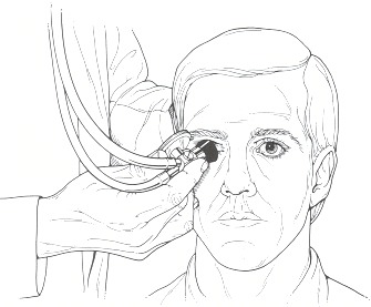
Figure 18.1
Technique for orbital auscultation.
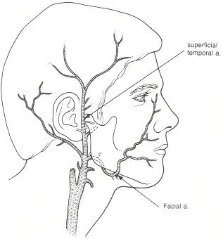
Figure 18.2
Arrows indicate points of manual compression of the right superficial temporal and facial arteries.
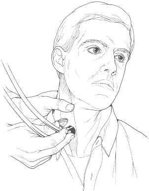
Figure 18.3
Technique for ausculting and abolishing venous hum.
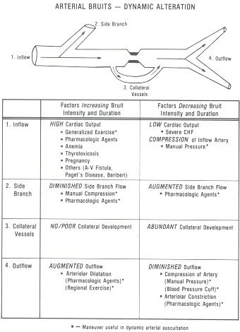
Figure 18.4
Summary of factors altering intensity and duration of arterial bruits. See text for details. A-V = arteriovenous; CHF = congestive heart failure. From Kurtz KJ. Dynamic vascular auscultation. Am J Med 1984;76:1066–74. Reproduced with permission.
Copyright © 1990, Butterworth Publishers, a division of Reed Publishing
Không có nhận xét nào:
Đăng nhận xét